

















7 Best CCK-8 Assay Kits for Cell Viability Testing
Cell viability measurements shape decisions in drug screening, toxicology, and routine cell culture. Cck-8 assays are popular because they produce a soluble color signal without a washing step. Yet a brighter well does not automatically mean more living cells. Cck-8 measures cellular metabolic activity, which can shift with cell density, treatment, and incubation time.
The wider cell-culture market provides useful context, but it is not a direct measure of CCK-8 kit sales. Grand View Research estimated the global cell-culture market at USD 27.9 billion in 2023 and projected continued growth through 2030. These figures describe a broad field, not assay-kit demand. That distinction matters. In the Assay Guidance Manual, cell-based assay scientist Terry Riss and co-authors write: “Cell viability assays are used to determine the number of living and/or dead cells in a sample.” Their definition highlights why assay choice and validation matter.
This guide compares seven CCK-8 assay kits by signal performance, protocol clarity, compatibility, and practical handling. It considers details that affect real bench work, such as edge-well variation, plate format, and reagent stability. A kit may look excellent on paper. Your cells may disagree. Results should be checked against appropriate controls and, where needed, an independent readout. No single kit suits every cell line or experimental question. The comparison is a starting point, not a substitute for validation in your own model.
How CCK-8 Assays Measure Cell Viability
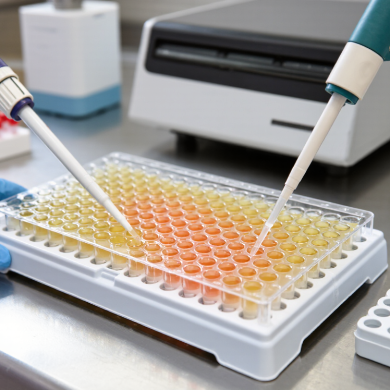
CCK-8 assays estimate cell viability through a color-producing chemical reaction. The reagent contains WST-8, a tetrazolium salt that living cells reduce through cellular dehydrogenase activity, with help from electron-transfer compounds. This reaction produces orange, water-soluble formazan in the culture medium. More color often indicates more metabolically active cells. Not a direct count. A plate reader measures absorbance, commonly near 450 nm.
The signal is useful, but it needs careful interpretation. Drug exposure, nutrient stress, or other metabolic changes can alter the color even when cell numbers barely shift. A stronger signal does not automatically mean more cells. Include untreated controls and reagent-only blanks to assess the response and background. Test a range of cell densities, too; crowded wells may saturate the signal, while sparse wells can sit near background. Keep incubation times consistent, since color can continue developing.
In practice, even seeding matters. Replicate wells can reveal uneven cell distribution, a small but common source of confusing results. Avoid bubbles in the optical path, and check that the chosen reading time fits the cells and experimental conditions. Interpret the measurement as metabolic activity under those conditions, rather than proof that every cell is alive. That distinction is easy to overlook.
